


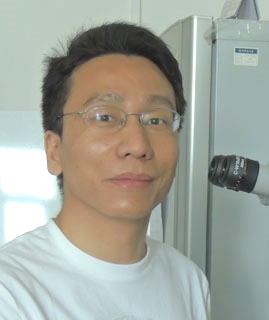
张少兵
教授,博士生导师
-
联系方式
Tel:010-68901204
Email: soz001@@cnu.edu.cn(去掉一个@)
北京市西三环北路105号首都师范大学生命科学学院
邮编100048 -
招生方向
博士:生物化学与分子生物学、分子信号传导
硕士:生物化学与分子生物学、脂代谢与疾病
研究概述/Research
脂滴是真核生物细胞储存脂肪的专职细胞器,脂滴的大小和数量直接决定细胞、组织、个体水平上的脂肪含量,脂滴代谢紊乱可导致肥胖症、二型糖尿病、心血管疾病等代谢疾病。脂滴生成、长大、融合、水解等动态过程的核心细胞生物学原理是什么(图1)?脂滴的大小和数量是如何被脂滴衣蛋白、代谢途径、信号通路综合调控的?这些问题迄今为止仍未得到很好的解答。
在研究一类过氧化物酶体功能缺失的秀丽线虫突变体的基础上,我们率先鉴定了线虫细胞里的脂肪存储结构是脂滴而非先前所认为的“类溶酶体”、“肠颗粒”或“不同于类溶酶体的膜泡”,厘清了线虫脂肪研究领域里概念和方法的错误,建立了一套正确的脂滴染色标记、体积测量、脂肪含量测定等方法(BMC Cell Biology 2010),并且,我们建立了第一例线虫脂滴大小调控的遗传学和细胞生物学模型(Proc. Natl. Acad. Sci. U. S. A. 2010)。本实验室运用这些研究方法,并穷尽强大的线虫分子遗传学,完成首例饱和正向遗传学筛选(图2),筛选到118个超大脂滴突变体,这些突变体被命名为drop (lipid droplet abnormal),归属于13个突变基因,分别显著上调脂滴长大、融合等过程,或显著下调脂滴水解过程(Genes Genomes Genetics 2016)。我们已经克隆了全部13个drop基因,并在特定的drop基因突变的背景上进行了增强子突变体和抑制子突变体筛选,又分离克隆了30个新的drop基因。我们正在进行分子遗传学、细胞生物学和脂质化学分析,力图深入理解drop基因编码的DROP蛋白调控脂滴长大、融合、水解的精微原理。
本实验室当前集中研究的一个过程是脂滴融合。我们发现:细胞色素P450蛋白CYP-37A1(DROP-1)和P450氧化还原酶EMB-8(DROP-8)共同合成一种脂溶性的激素,该激素抑制核受体DAF-12(DROP-31);当CYP-37A1失去功能后,激素不能合成,DAF-12促进肠道细胞里发生热敏型脂滴融合,热敏型脂滴融合类似于哺乳动物褐色脂肪细胞转化为白色脂肪细胞时脂滴的形态变化(Proc. Natl. Acad. Sci. U. S. A. 2017)。后续研究发现,当CYP-37A1失去功能后,DAF-12上调溶血磷脂酰胆碱酰基转移酶MBOA-6(DROP-33)和包括FAT-3(DROP-35)在内的7个脂肪酸去饱和酶的转录和脂滴定位,这些酶在脂滴表面合成富含ω-3 20碳多不饱和脂肪酸的磷脂酰胆碱,从而升高磷脂单分子层形成的脂滴膜的流动性,促进热敏型融合(Nature Communications 2025)(图3)。CYP-37A1、EMB-8、DAF-12、MBOA-6、FAT-3在人体内的同源蛋白分别是CYP4V2、POR、FXR、LPCAT3、 FADS2,我们发现人CYP4V2、POR、LPCAT3具有保守的调控脂滴大小和脂滴融合的功能。
我们所建立的线虫脂滴长大和融合模型既能快捷地结合运用遗传学、细胞生物学、生物化学、脂类分析化学等技术手段,又能有效整合分子、细胞、组织、个体水平上的研究结果,这些模型让我们揭示了脂滴细胞生物学过程的一些核心保守原理。我们期待线虫脂滴研究为哺乳动物和人类脂滴研究提供可以借鉴的模式,帮助找出让人罹患脂代谢疾病的遗传和饮食因素,并协助预防诊断、开发新药和疗法。
Lipid droplets are cellular organelles dedicated for fat storage and fat mobilization in eukaryotic cells. The size and number of lipid droplets directly determine fat levels of cells, tissues, and organisms. Disorders of lipid droplet metabolism may lead to obesity, type 2 diabetes, cardiovascular disease, and other metabolic diseases. What are the core mechanisms underlying the dynamic cellular processes of lipid droplets such as de novo formation, growth, fusion, and hydrolysis (Fig. 1)? How are lipid droplet size and number integratively regulated by lipid droplet coat proteins, metabolic pathways, and signaling proteins? These questions are poorly understood.
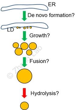
Fig. 1. Basic cellular processes of lipid droplets (LD, golden): de novo formation, growth, fusion, and hydrolysis. Green arrows indicate anabolic processes; red, catabolic. The linear order of these processes is not necessarily true. Little is known about the morphological intermediates and molecular and cellular mechanisms of these processes.
In studying a class of peroxisomal fatty acid β-oxidation mutants, we discovered that the cellular fat storage structures in the model animal Caenorhabditis elegans are lipid droplets but not “lysosome-related organelles (LROs)”, “gut granules”, or the so called “vesicles distinct from LROs”. This study thus cleared conceptual confusions and technical mistakes off the C. elegans fat research field, and established a suite of correct lipid droplet staining and marking, size measurement, fat level measurement methods (BMC Cell Biology 2010). Furthermore, we established the first genetic and cell biological model of C. elegans lipid droplet size regulation (Proc. Natl. Acad. Sci. U.S.A. 2010). By exploiting these methods and exhausting the awesome power of C. elegans molecular genetics, our laboratory conducted and reported the first saturated forward genetic screen for mutants with supersized lipid droplets (Fig. 2). We isolated 118 supersized lipid droplet mutants and named these mutants as drop (lipid droplet abnormal). These mutants belong to 13 mutant drop genes, which significantly up-regulate lipid droplet growth or lipid droplet fusion, or significantly down-regulate lipid droplet hydrolysis (Genes Genomes Genetics 2016). We have cloned all 13 drop genes. We have also conducted enhancer and suppressor mutant screens in the background of certain drop gene mutants, and have cloned nearly 30 new drop genes. We are carrying out molecular genetics, cell biology, and lipid analytical chemistry analyses with these genes. We aim to uncover the intricate mechanisms by which the DROP proteins encoded by drop genes regulate the growth, fusion, and hydrolysis of lipid droplets.
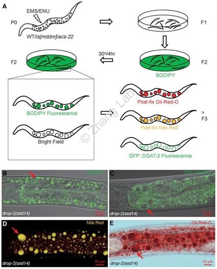
Fig. 2. A forward genetic screen for C. elegans mutants with supersized lipid droplets. (A) Mutagens (EMS and ENU) were used to mutagenize C. elegans of P0 generation. F2 progeny were vital stained with BODIPY dye. Candidate mutants were isolated by virtue of supersized BODIPY-positive and light-refracting globular structures. Progeny (>F3) of F2 isolates were then tested by postfix Oil-Red-O staining, postfix Nile Red staining, and GFP::DGAT-2 marking. (B-D) Supersized lipid droplets (arrows) in drop-2(ssd14), one of the 118 mutant isolates, were readily visible in bright field and were labeled by vital BODIPY (B), by GFP::DGAT-2 (C), by postfix Nile Red with a true color of gold (D), and by postfix Oil-Red-O (E). Modified from Genes Genomes Genetics 2016.
One of our current projects is focused on lipid droplet fusion. We discovered that cytochrome P450 protein CYP-37A1 (DROP-1) and P450 oxidoreductase EMB-8 (DROP-8) act together to synthesize a putative lipophilic hormone. This hormone inhibits the nuclear hormone receptor DAF-12 (DROP-31). When CYP-37A1 is dysfunctional, the hormone is not synthesized so that DAF-12 becomes active to promote thermosensitive lipid droplet fusion in intestine cells. The thermosensitive lipid droplet fusion is morphologically analogous to what happens to lipid droplets in the conversion of brown/beige adipocytes to white adipocytes in mammals (Proc. Natl. Acad. Sci. U. S. A. 2017). Subsequent research reveals that in cyp-37A1 mutants, DAF-12 up-regulates the transcription and LD targeting of lysophosphatidylcholine acyltransferase MBOA-6 (DROP-33) and of seven fatty acid desaturases including FAT-3 (DROP-35). These enzymes act on the surface of lipid droplets to synthesize phosphatidylcholine containing ω-3 C20 polyunsaturated fatty acids (ω-3 C20-PC). The high level of ω-3 C20-PC increases the fluidity of the mono-layered lipid droplet membrane to promote thermosensitive fusion (Nature Communications 2025) (Fig. 3). The homologs of CYP-37A1, EMB-8, DAF-12, MBOA-6, and FAT-3 in humans are CYP4V2, POR, FXR, LPCAT3, and FADS2, respectively. We found that CYP4V2, POR, and LPCAT3 also play roles in lipid droplet size regulation and or lipid droplet fusion.
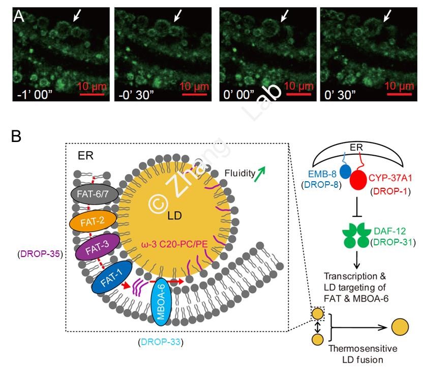
Fig. 3. Specific regulation of thermosensitive lipid droplet fusion by the CYP-37A1-EMB-8-hormone-DAF-12 pathway in C. elegans. (A) In the cyp-37A1 (drop-1) mutant, two lipid droplets (labeled by GFP::DGAT-2) fuse into a supersized one (arrow) at the high temperature of 30 oC. The fusion is direct membrane fusion, displays no asymmetrical partnership, and finishes in seconds. (B) The thermosensitive lipid droplet fusion is repressed in wild-type animals by cytochrome P450 protein CYP-37A1 (DROP-1) and P450 oxidoreductase EMB-8 (DROP-8), which together synthesizes a putative hormone. The hormone represses the transcriptional activity of nuclear hormone receptor DAF-12 (DROP-31). In cyp-37A1 mutants, DAF-12 is relieved to up-regulate the transcription and lipid droplet targeting of fatty acid △6 desaturase FAT-3 (DROP-35), other FAT desaturases, and lysophosphatidylcholine acyltransferase MBOA-6 (DROP-33). These enzymes work in relay to synthesize ω-3 C20 polyunsaturated fatty acid-containing phosphatidylcholine(ω-3 C20-PC)for lipid droplet membrane. The elevated level of ω-3 C20-PC increases the fluidity of lipid droplet membrane, which promotes thermosensitive lipid droplet fusion. Adapted from Genes Genomes Genetics 2016, Proc. Natl. Acad. Sci. U. S. A. 2017, and Nature Communications 2025.
The C. elegans lipid droplet models that we have established lend us two advantages: a combination of molecular genetics, cell biology, biochemistry, and lipid analytical chemistry, and, an integration of results obtained at the levels of molecules, cells, tissues, and whole animals. These models have allowed us to reveal some of the conserved mechanisms of lipid droplet processes. We expect to present a C. elegans paradigm of lipid droplet regulation to benefit the research of mammalian and human lipid droplets, with an ultimate goal of identifying genetic and dietary risk factors, prevention guidelines, and therapeutic interventions for human metabolic diseases.
发表论文/Publications
Wang X, Qiu Y, Niu H, Liu M, Tan S, Zhang SO (2026) A new set of combinatorial dimeric codes for nuclear hormone receptor-target gene specificity. Nucleic Acids Research 54: gkag058.
Li Q, Zhou X, Zhang X, Zhang C, Zhang SO (2025) Nuclear receptor signaling regulates compartmentalized phosphatidylcholine remodeling to facilitate thermosensitive lipid droplet fusion. Nature Communications 16: 3955.
Zhang YP, Zhang WH, Zhang P, Li Q, Sun Y, Wang JW, Zhang SO, Cai T, Zhan C, Dong MQ (2022) Intestine-specific removal of DAF-2 nearly doubles lifespan in Caenorhabditis elegans with little fitness cost. Nature Communications 13: 6339.
Gao C, Li Q, Yu J, Li S, Cui Q, Hu X, Chen L, Zhang SO (2021) Endocrine pheromones couple fat rationing to dauer diapause through HNF4α nuclear receptors. Science China Life Sciences 64: 2153-2174. (Commented in Science China Life Sciences; reported by EurekAlert!, Mirage News, and Science X )
Li S, Li Q, Kong Y, Wu S, Cui Q, Zhang M, Zhang SO (2017) Specific regulation of thermosensitive lipid droplet fusion by a nuclear hormone receptor pathway. Proc. Natl. Acad. Sci. U. S. A. 114: 8841-8846.
Li S, Xu S, Ma Y, Wu S, Feng Y, Cui Q, Chen L, Zhou S, Kong Y, Zhang X, Yu J, Wu M, Zhang SO (2016) A Genetic Screen for Mutants with Supersized Lipid Droplets in Caenorhabditis elegans. G3: Genes Genomes Genetics 6: 2407-2419. (Paper downloaded the most from the journal in the month; recommended as Paper of the Month by Genetics Society of America)
Zhang P, Na H, Liu Z, Zhang S, Xue P, Chen Y, Pu J, Peng G, Huang X, Yang F, Xie Z, Xu T, Xu P, Ou G, Zhang SO*, Liu P* (2012) Proteomic study and marker protein identification of Caenorhabditis elegans lipid droplets. Molecular & Cellular Proteomics 11: 317-328. (* co-corresponding author)
Xu N, Zhang SO, Cole RA, McKinney SA, Guo F, Haas JT, Bobba S, Farese RV Jr, Mak HY (2012) The FATP1-DGAT2 complex facilitates lipid droplet expansion at the ER-lipid droplet interface. Journal of Cell Biology 198: 895-911. (Cover article; recommended by Faculty of 1000 Biology)
Zhang SO, Trimble R, Guo F, Mak HY (2010) Lipid droplets as ubiquitous fat storage organelles in C. elegans. BMC Cell Biology 11: 96. (Rated as Highly Accessed & Featured by BMC Cell Biology)
Zhang SO, Box A, Xu N, Le Men J, Yu J, Guo F, Trimble R, Mak HY (2010) Genetic and dietary regulation of lipid droplet expansion in Caenorhabditis elegans. Proc. Natl. Acad. Sci. U. S. A. 107: 4640-4645. (Recommended by Faculty of 1000 Biology)
Zhang SO, Mathur S, Hattem G, Tassy O, Pourquié O (2010) Sex-dimorphic gene expression and ineffective dosage compensation of Z-linked genes in gastrulating chicken embryos. BMC Genomics 11: 13.
Zhang SO, Kuo D-H, Weisblat DA (2009) Grandparental stem cells in leech segmentation: Differences in CDC42 expression are correlated with an alternating pattern of blast cell fates. Developmental Biology 336: 112-131.
Zhang SO, Weisblat DA (2005) Applications of mRNA injections for analyzing cell lineage and asymmetric cell divisions during segmentation in the leech Helobdella robusta. Development 132: 2103-2113.
加入我们/Join
本实验室研究组长 2005年博士毕业于美国加州大学伯克利分校(Berkeley),2006-2011年先后在美国霍华德休斯医学研究所(HHMI)、斯陶尔思医学研究所(Stowers)从事博士后研究。自2011年在首都师大建立实验室以来,我们利用遗传学、细胞生物学、生物化学、显微成像及其它必要的方法手段,系统地研究调控脂滴动态过程的遗传通路和核心生化原理。我们欢迎踏实勤奋的学生来本实验室研修博士学位,我们也欢迎新近的博士毕业生来本实验室从事博士后训练。优秀的博士生将会有机会被送到国内外知名高校进行交流培养、参加国际学术会议。感兴趣的个人请提前联系组长,组长将根据候选人的具体情况做出录用意向。